44 adipose tissue with labels
Adipose the Label Adipose the Label. Bright colours and prints, in silhouettes that expand or enhance the attitude of the wearer not for flattering the figure. The Beauty of Adipose Inhibition. Disengage. Liberation. Quick links. Search Info. Search Our mission. Adipose The Label is about body positive attitudes that show personality and has no care for how a ... Evaluation of Clinical Efficacy of Autologous Adipose Tissue Derived ... This is prospective, open label, single arm clinical study that will establish the safety and efficacy of adipose derived stromal cells in patients with osteoarthritis disease. Total 140 subjects were enrolled in the study. Each subject will receive intra-articular Autologous ADSC with PRP along with standard treatment for osteoarthritis disease.
BIOMIMESYS® Adipose tissue - HCS Pharma BIOMIMESYS® Adipose tissue. BIOMIMESYS® is a unique groundbreaking 3D cell culture technology which associates the behavior of a solid scaffold and of a hydrogel. It provides a cell culture microenvironment reproducing all aspects of human tissues, including matrix architecture, cellular organization, cell-cell and cell-matrix interactions ...
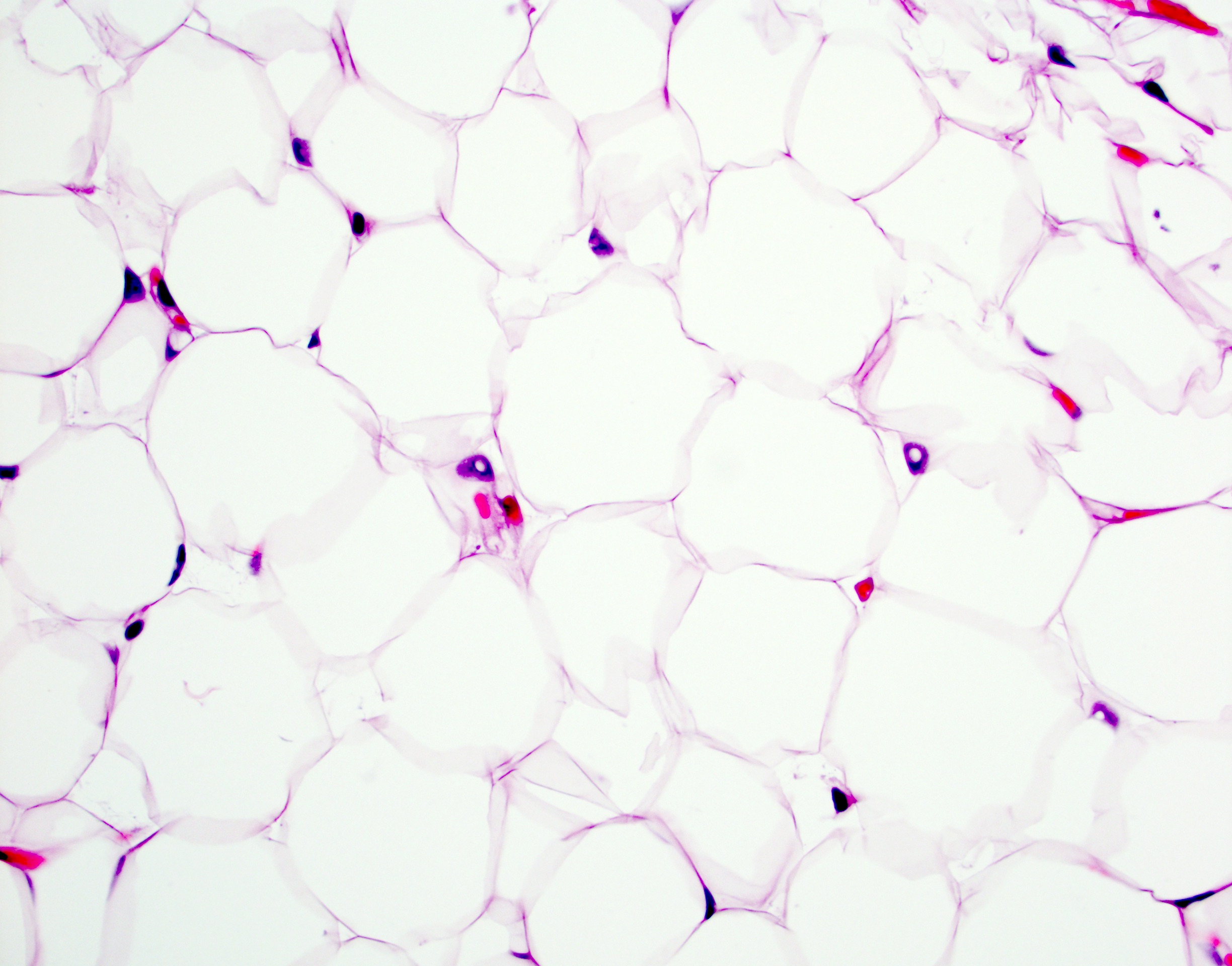
Adipose tissue with labels
Adipose Tissue Anatomy | Plastic Surgery Key Adipose Tissue Anatomy. Abdominal WAT: the tissue is characterized by large adipose cells and poor collagenic component. ( a) Light microscopy (scale bar, 15 μm). ( b) SEM (scale bar, 50 μm). ( c) SEM (scale bar, 20 μm). The adipocytes are surrounded by a thin capsule of collagen fibers. ( d) TEM (scale bar, 2,000 μm). Quantum dots for labeling adipose tissue-derived stem cells Adipose tissue-derived stem cells (ASCs) have a self-renewing ability and can be induced to differentiate into various types of mesenchymal tissue. Because of their potential for clinical application, it has become desirable to label the cells for tracing transplanted cells and for in vivo imaging. … Two-photon excited fluorescence of intrinsic fluorophores enables label ... Adipose tissue function has been recognized to exert significant influence on systemic metabolic balance and overall homeostasis health through energy storage via lipid accumulation, direct energy expenditure through substrate oxidation 1,2, and secretion of various signaling and regulatory molecules 3,4.Major health problems such as type 2 diabetes mellitus, cancer, and cardiovascular disease ...
Adipose tissue with labels. Label-free profiling of white adipose tissue of rats exhibiting high or ... We analysed visceral adipose tissue of HCR and LCR using label-free HDMS E profiling. The running capacity of HCR was 9-fold greater than LCR. Proteome profiling encompassed 448 proteins and detected 30 significant (p <0.05; false discovery rate <10%, calculated using q-values) differences. Approximately half of the proteins analysed were of ... Adipose tissue - Vida Private Label Our formulations, which are designed to work on adipocytes, are based on active ingredients containing xanthines, such as caffeine and theophylline, known for their beneficial remodelling action on fat cells, and sodium deoxycholate. Adipose Tissue: What Role Does It Play In Diabetes? - Ben's Natural Health An Adipose Tissue Mass connects organs and tissues and is an insulator against cold. When these tissues form, new blood vessels form from existing ones. The splitting of the vascular growth causes tissue dysfunction. ... Understand what you eat-read food labels; Reduce the amount of added sugar intake (Men <9tsp/day, women<6 tsp) Live and ... Adipose Tissue: What Is It, Location, Function, and More - Osmosis Adipose tissue is a specialized type of connective tissue that arises from the differentiation of mesenchymal stem cells into adipocytes during fetal development. Mesenchymal stem cells are pluripotent cells that can transform into various cell types, including fat cells, bone cells, cartilage cells, and muscle cells, among others.
The developmental origins of adipose tissue | Development | The Company ... In mammals, adipose tissue forms in utero, in the peripartum period and throughout life.Notably, even in adult humans new adipocytes are generated continually and at substantial rates (Spalding et al., 2008).Adipose tissue is composed of adipose stem cells (the precursor cells that give rise to new adipocytes), adipocytes (the fat-storing cells) and various other cell types, which include ... Labeled Tissues Flashcards | Quizlet Blood cells. Label ground substance, elastic fibers, collagen fibers, fat droplet, specific cells of each tissue. Nervous Tissue. Label Cell Body, nucleus and nerve fibers ( cell extensions. Simple Cuboidal Epithelium. Label Free surface, nuclei, and basement membrane. Simple Columnar Epithelium. Label Free surface, nuclei, and basement membrane. Adipose Tissue - Composition, Location and Function - ThoughtCo Adipose Tissue Location . Adipose tissue is found in various places in the body. Some of these locations include the subcutaneous layer under the skin; around the heart, kidneys, and nerve tissue; in yellow bone marrow and breast tissue; and within the buttocks, thighs, and abdominal cavity. While white fat accumulates in these areas, brown fat is located in more specific areas of the body. Adipose Tissue: Types and Location - Study.com Cell-signaling adipokines in the adipose tissue send messages to and from other organs of the body to regulate energy balance, inflammation, hunger and satiety, insulin sensitivity, and cell ...
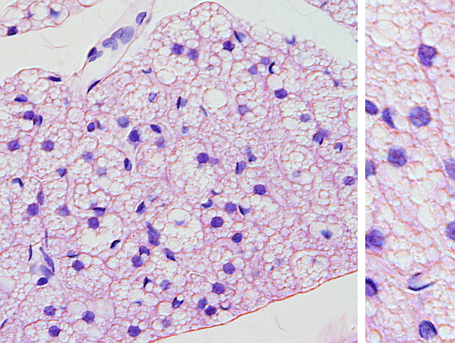
ANATOMY TISSUES LABELING Flashcards | Quizlet adipose tissue. loose areolar connective tissue. hyaline cartilage. compact bone. hyaline cartilage. dense regular connective tissue. adipose tissue. blood. compact bone. stratified squamous epithelium. simple ciliated columnar epithelium. pseudostratified ciliated columnar epithelium. Brown Adipocytes - Yale University Brown adipose tissue is typically found in large amounts in newborns and some hibernating animals and is important as a source of energy. The cells in brown fat contain numerous and very distinct lipid droplets. The presence of an uncoupling protein in these cells causes the generation of heat that allows for non-shivering thermogenesis. Adipose Tissue Histology Brown adipose tissue (labels) - histology slide: Adipose tissue, mouse - histology slide: Adipose tissue - histology slide: Adipose tissue - histology slide: ... Adipose tissue - histology slide: Adipose tissue - histology slide: Adipose tissue - histology slide: Adipose tissue - histology slide: 27 files on 3 page(s) 1: 2: 3: Powered by ... 260 Adipose tissue microscope Images, Stock Photos & Vectors | Shutterstock Find Adipose tissue microscope stock images in HD and millions of other royalty-free stock photos, illustrations and vectors in the Shutterstock collection. Thousands of new, high-quality pictures added every day.
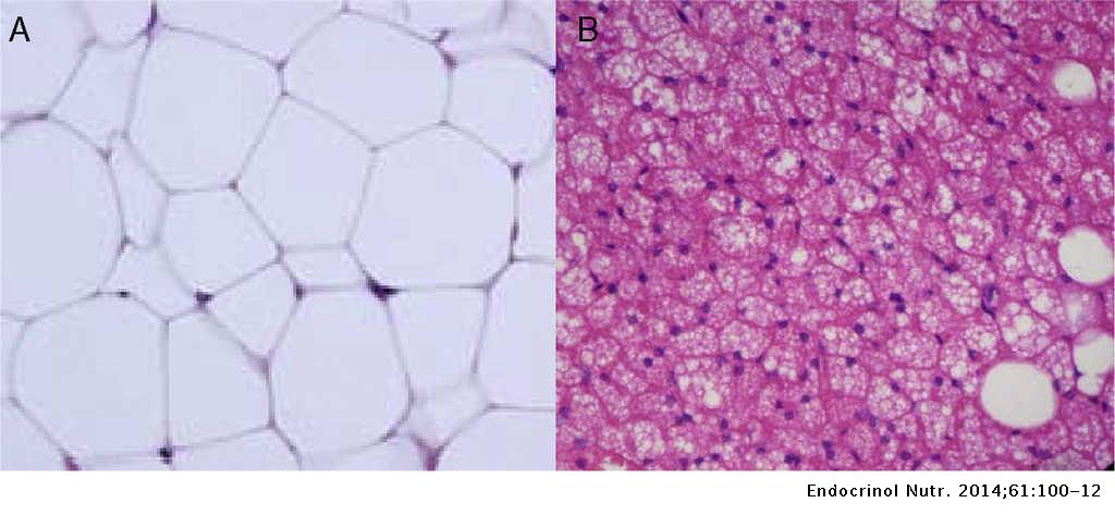
Adipose Tissue - The Definitive Guide| Biology Dictionary Adipose tissue is split into two main types of connective tissue - white and brown - that store and burn energy respectively. ... In this heat-generating response, UCP 1 - the picture below labels it thermogenin - the purple oval in the gray mitochondria - is released from the mitochondria of the brown adipose tissue. This protein ...
Solved Label the components of adipose tissue. Fibroblast - Chegg Expert Answer. Answer The label is indicated from TOP to …. View the full answer. Transcribed image text: Label the components of adipose tissue. Fibroblast Cell membrane Elastic fiber Nucleus Adipocyte.
Skin and hypodermis - Eugraph The skin consists of two layers, the epidermis and the dermis. Below the skin is a layer of areolar/adipose tissue called the hypodermis (or subcutaneous layer). The 40X image shows: e = epidermis. d = dermis. h = hypodermis. sw = sweat gland. seb = sebaceous gland. hf = hair follicle. p = papillary layer of dermis. r = reticular layer of dermis
Adipose tissue: Definition, location, function | Kenhub 1/2. Adipose tissue is a specialized connective tissue consisting of lipid-rich cells called adipocytes. As it comprises about 20-25% of total body weight in healthy individuals, the main function of adipose tissue is to store energy in the form of lipids (fat). Based on its location, fat tissue is divided into parietal (under the skin) and ...
Adipose Tissue - Meaning, Types, Functions, and FAQs - VEDANTU An Introduction. Adipose tissue is a complex, essential, and highly active metabolic and endocrine organ. It is a source of several hormones, including leptin, estrogen and resistin. Adipose tissue is composed primarily of adipocytes or fat cells. These comprise lipid storage droplets, which contain triacylglycerol and vary in size depending on ...
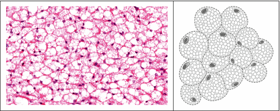
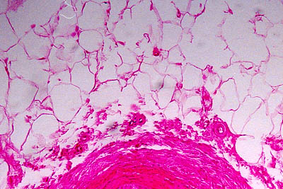
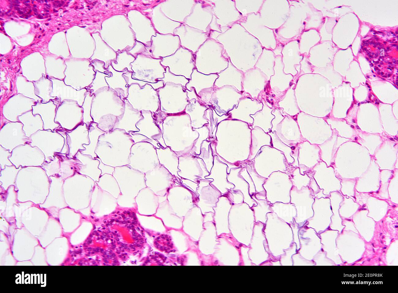
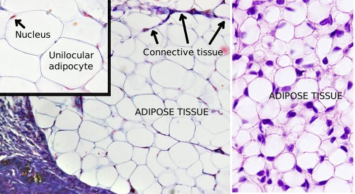
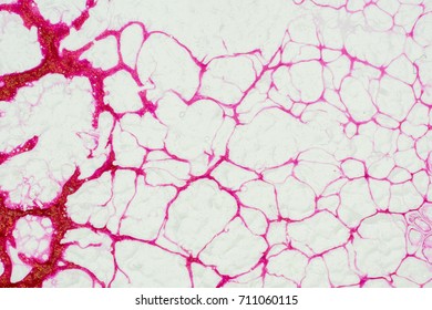
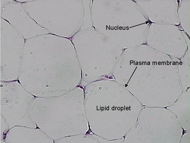
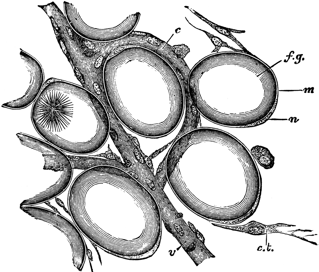
Adipose Issue - University of Wisconsin-La Crosse 1. Cell membrane. 2. Cell nucleus. 3. Fat vacuoles. Adipose connective tissue cells are specialized for fat storage and do not form ground substance or fibers. On prepared slides, adipose tissue appears somewhat like a fish net with white spaces connected together in a network. The cytoplasm and nucleus have been pushed to one side by a single ...
Adipose tissue - Wikipedia In humans, adipose tissue is located: beneath the skin (subcutaneous fat), around internal organs (visceral fat), in bone marrow (yellow bone marrow), intermuscular (Muscular system) and in the breast (breast tissue).Adipose tissue is found in specific locations, which are referred to as adipose depots.Apart from adipocytes, which comprise the highest percentage of cells within adipose tissue ...
Solved Label the parts of the skin. Adipose tissue Stratum - Chegg Question: Label the parts of the skin. Adipose tissue Stratum basale Dermal papilla Stratum corneum Stratum spinosum Basement membrane Sebaceous gland Hair shaft Muscle layer Hair follicle Reset Zoom . This problem has been solved! See the answer See the answer See the answer done loading.
Open Label, Multi-Center Study, Evaluating the Effect of Adipose Tissue ... This is an open label multi-center study with the aim of evaluating the efficacy of adipose tissue processed with the SyntrFuge™ system in facial aesthetics and contouring. Patients will be enrolled to the treatment group with adipose tissue processed with the SyntrFuge™ system followed by an injection of autologous microsized adipose ...
Adipose connective tissue - austincc.edu Adipose connective tissue 400X. The bar labeled "a" indicates the width of one adipose cell (adipocyte). The light purple dots you see inside the cells are an artifact of process used to make the images, and do not represent real structures. The arrow points to the nucleus of one adipocyte. The nucleus and cytoplasm are pushed to the outside of ...
Adipose tissue aging: mechanisms and therapeutic implications In an open-label phase 1 pilot study of Senolytics, old patients with diabetic kidney disease treated with Senolytics showed a reduction in adipose tissue senescent cell burden and key SASP ...
Off-label use of adipose-derived stem cells - PubMed The ease of cell harvest and high yield with minimal donor-site morbidity makes adipose tissue an ideal source of stem cells. Further, the multi-lineage potential of these cells present significant opportunities within the field of tissue engineering, with studies successfully demonstrating their ability to produce a range of tissue types.
Two-photon excited fluorescence of intrinsic fluorophores enables label ... Adipose tissue function has been recognized to exert significant influence on systemic metabolic balance and overall homeostasis health through energy storage via lipid accumulation, direct energy expenditure through substrate oxidation 1,2, and secretion of various signaling and regulatory molecules 3,4.Major health problems such as type 2 diabetes mellitus, cancer, and cardiovascular disease ...
Quantum dots for labeling adipose tissue-derived stem cells Adipose tissue-derived stem cells (ASCs) have a self-renewing ability and can be induced to differentiate into various types of mesenchymal tissue. Because of their potential for clinical application, it has become desirable to label the cells for tracing transplanted cells and for in vivo imaging. …
Adipose Tissue Anatomy | Plastic Surgery Key Adipose Tissue Anatomy. Abdominal WAT: the tissue is characterized by large adipose cells and poor collagenic component. ( a) Light microscopy (scale bar, 15 μm). ( b) SEM (scale bar, 50 μm). ( c) SEM (scale bar, 20 μm). The adipocytes are surrounded by a thin capsule of collagen fibers. ( d) TEM (scale bar, 2,000 μm).


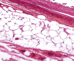






:watermark(/images/watermark_only.png,0,0,0):watermark(/images/logo_url.png,-10,-10,0):format(jpeg)/images/anatomy_term/adipocytes-3/XJcofGYn074KZFZBhpkSpQ_Adipocytes.png)

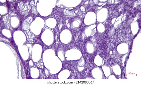
![HLS [ Connective Tissue, unilocular (white) adipocytes] HIGH ...](https://www.bu.edu/phpbin/medlib/histology/i/08202loa.jpg)




Post a Comment for "44 adipose tissue with labels"