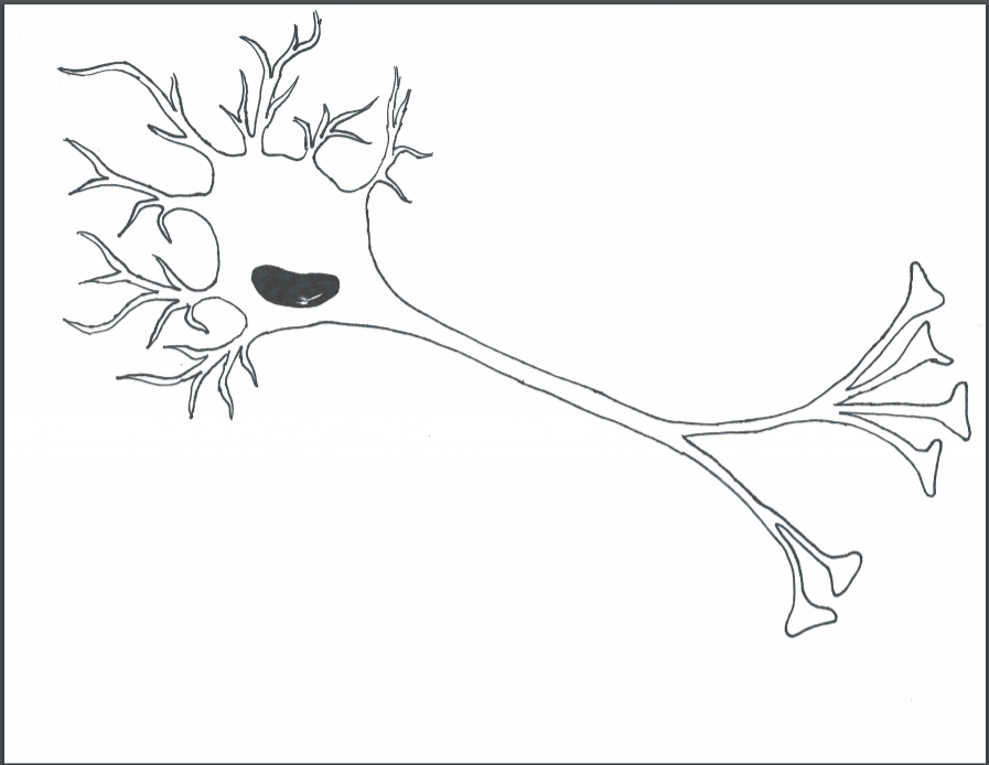
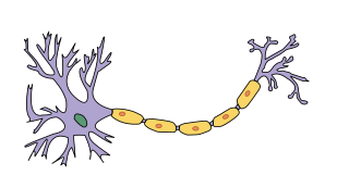
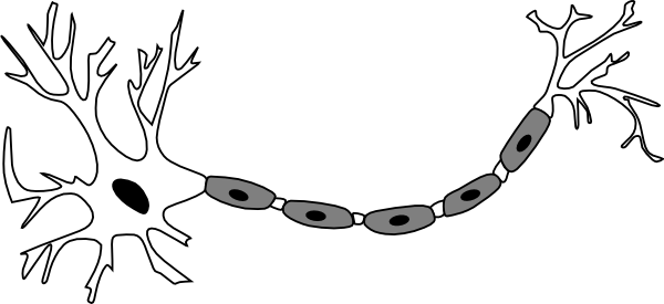
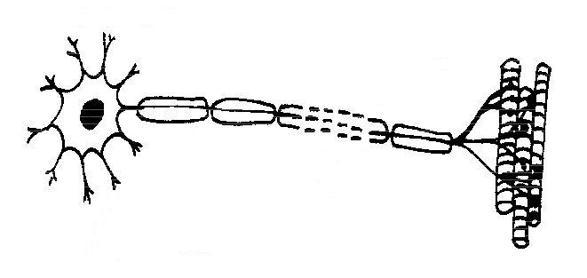
43 picture of a neuron without labels
how to draw structure of neuron/neuron diagram labelled ... Neuron Diagram Printable Worksheet. Description This printable 2-pack features a high-quality neuron diagram and is ideal for quizzes, homework, review work, ... Draw the diagram of neuron and label any two parts. Draw the diagram of neuron and label any two parts. · Neurons: Structure and Types · Synaptic Transmission of Nerve Impulse · Generation and Conduction of Nerve ...
100+ Free Neuron & Brain Images - Pixabay 194 Free images of Neuron. Related Images: neurons brain network nervous system mind mental health psychology biology web ...
Picture of a neuron without labels
Picture of a neuron without labels Picture of a neuron without labels alexandra daddario hot one the woman Interneurons connect various neurons within the brain and spinal cord. The simplest type of neural pathway is a monosynaptic (single connection) reflex pathway, like the knee-jerk reflex. MonkeyLogic - Brown University Introduction. MonkeyLogic is a MATLAB toolbox for the design and execution of psychophysical tasks with high temporal precision.It is structured to allow for the flexible construction of sensory, motor, or cognitive tasks that are based upon the interaction of a subject with visual stimuli through the use of eye-position, joystick, button, lever, and / or keyboard input. Photo-labeling neurons in the Drosophila brain - ScienceDirect The cell body of the neuron of interest cannot be located before starting the photo-labeling procedure (step 2 of the photo-label individual neurons section). Potential solution. If the cell body of the neuron of interest is located in the center of the brain, it may be difficult to image.
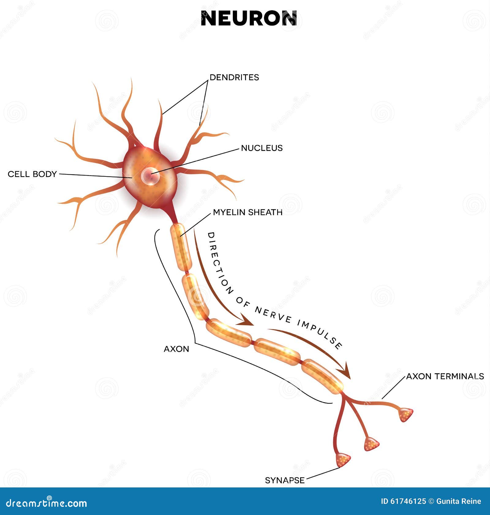
Picture of a neuron without labels. Labeled Neuron Diagram | Science Trends Neurons are a type of cell and are the fundamental constituents of the nervous system and brain. Neurons take in stimuli and convert them to electrical and chemical signals that are sent to our brain. There are 3 major kinds of neurons in the spinal cord: sensory, motor, and interneurons. Superstar Worksheets - Superstar Worksheets VerkkoFree Worksheets PRESCHOOL WORKSHEETS ⭐ ⭐ KINDERGARTEN WORKSHEETS ⭐ Alphabet WorksheetsFree Alphabet Letter Printables, ABC Tracing, Blank Templates. Coloring Pages, Mazes, Worksheets, Activities.... Math WorksheetsAddition, Multiplication, Place Value, Fractions, Number Order, Rounding, Comparing, Measurement The Neuron - BrainFacts The neuron is the basic working unit of the brain, a specialized cell designed to transmit information to other nerve cells, muscle, or gland cells. Neurons are cells within the nervous system that transmit information to other nerve cells, muscle, or gland cells. Most neurons have a cell body, an axon, and dendrites. Photo-labeling neurons in the Drosophila brain - PMC The entire morphology of the photo-labeled neuron is clearly distinguishable from other neurons that also express PA-GFP, but were not photo-labeled (gray arrow). The morphological features of the photo-labeled neuron — such as its axonal projections and presynaptic boutons (yellow arrows) are clearly visible.
Types of Neurons: Parts, Structure, and Function - Verywell Health Summary. Neurons are responsible for transmitting signals throughout the body, a process that allows us to move and exist in the world around us. Different types of neurons include sensory, motor, and interneurons, as well as structurally-based neurons, which include unipolar, multipolar, bipolar, and pseudo-unipolar neurons. An Easy Guide to Neuron Anatomy with Diagrams - SimplyPsychology.org The central nervous system, which comprises the brain and spinal cord, and the peripheral nervous system, which consists of sensory and motor nerve cells all contain these information processing neurons.. Parts of a Neuron. The neuron contains the soma (cell body) from which extend the axon (a nerve fiber conducting electrical impulses away from the soma) and dendrites (tree-like structures ... 2,834 Labeled brain anatomy Images, Stock Photos & Vectors - Shutterstock Find Labeled brain anatomy stock images in HD and millions of other royalty-free stock photos, illustrations and vectors in the Shutterstock collection. Thousands of new, high-quality pictures added every day. Michael Bolton - Wikipedia VerkkoMichael Bolotin (born February 26, 1953), known professionally as Michael Bolton, is an American singer and songwriter.Bolton originally performed in the hard rock and heavy metal genres from the mid-1970s to the mid-1980s, both on his early solo albums and those he recorded as the frontman of the band Blackjack.He became better known for …
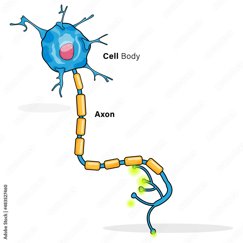
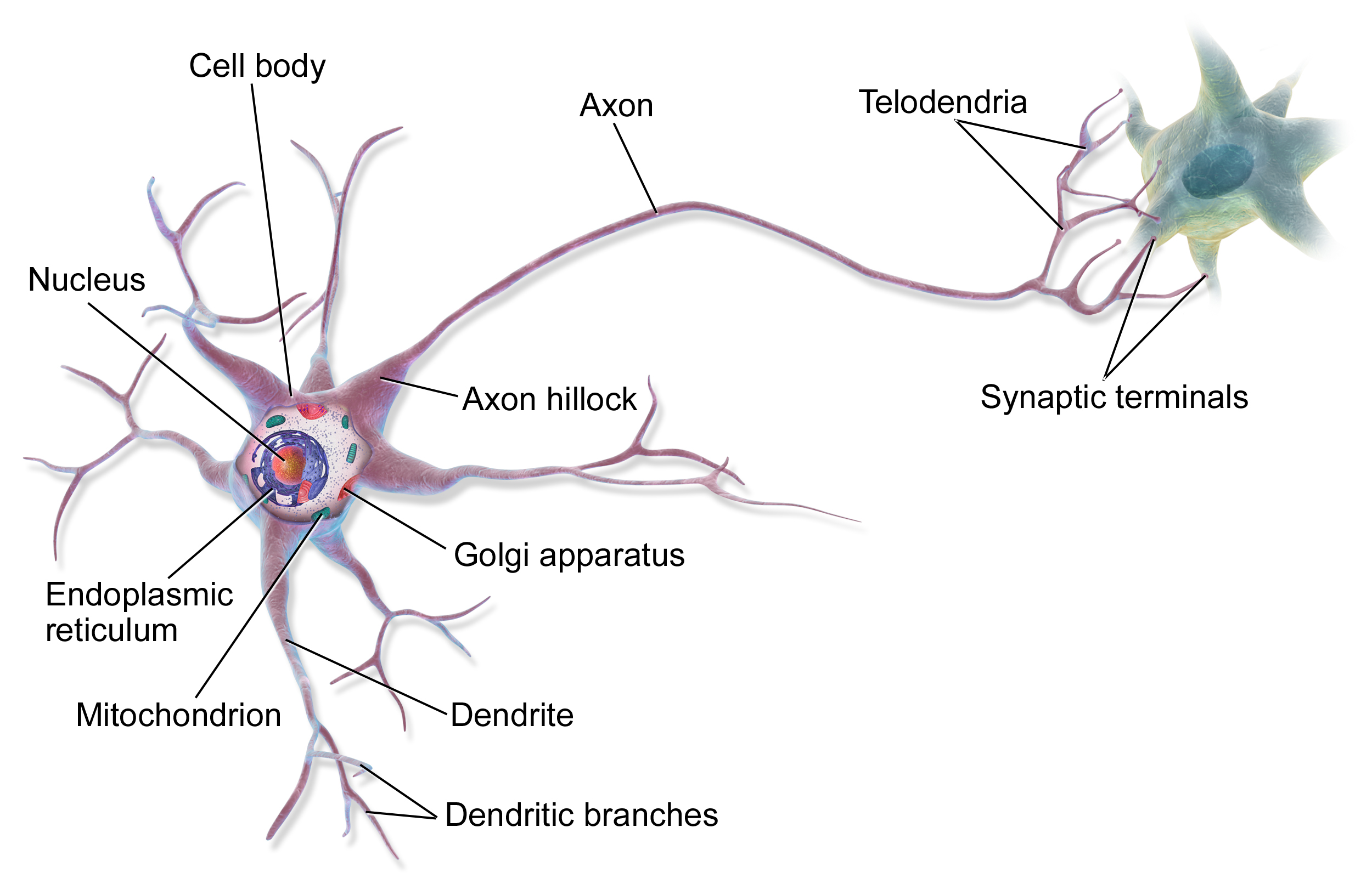
Neuron Label - Pinterest Nov 16, 2017 - This picture of the neuron is unlabeled, write in the labels to test your knowledge of the anatomy of a neuron. GitHub - andrewekhalel/MLQuestions: Machine Learning and … VerkkoSo we need to find the right/good balance without overfitting and underfitting the data. 2 ) What ... This is just finding how many input nodes actually connect through to a neuron in a ... PCA ignores class labels. We can picture PCA as a technique that finds the directions of maximal variance. In contrast to PCA, LDA attempts to find a ... A Guide to Understand Neuron with Neuron Diagram 3.1 How to Draw a Neuron Diagram from Sketch Step 1: First, the students need to draw a circle. Based on it, they need to draw a star-like shape. It is called the cell body of the neurons. One corner of the stars is extended, forming a very thin-tube-like structure-the Axon. American Express Picture of a neuron without labels Neurons, also known as nerve cells, are essentially the cells that make up the brain and the nervous system. Neurons do not touch each other, but where one neuron comes close to another neuron, a synapse is formed between the two.
Chapter 2 Test Review
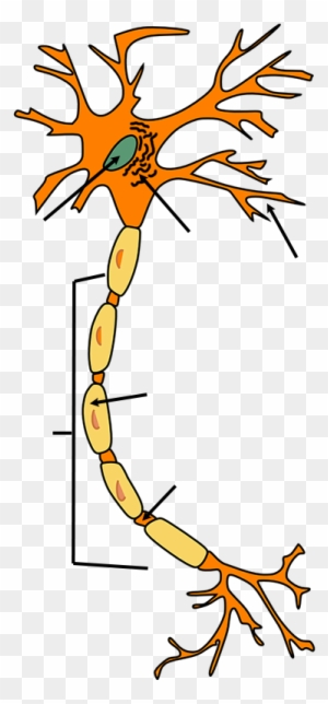
File:Derived Neuron schema with no labels.svg English: Diagram of neuron with arrows but no labels. ... You may do so in any reasonable manner, but not in any way that suggests the ...
Neuron Structure" Images – Browse 13 Stock Photos, Vectors ...
A Labelled Diagram Of Neuron with Detailed Explanations Diagram Of Neuron A neuron is a specialized cell, primarily involved in transmitting information through electrical and chemical signals. They are found in the brain, spinal cord and the peripheral nerves. A neuron is also known as the nerve cell.

File:Neuron (PSF).png - Wikimedia Commons
American Express The image_batch is a tensor of the shape (32, 180, 180, 3). This is a batch of 32 images of shape 180x180x3 (the last dimension refers to color channels RGB). The label_batch is a tensor of the shape (32,), these are corresponding labels to the 32 images.You can call .numpy () on the image_batch and labels_batch tensors to convert them to a.

Label the parts of a neuron in the following diagram ...
6.5.2 Draw and label a diagram of the structure of a motor neuron 6.5.2 Draw and label a diagram of the structure of a motor neuron. Watch later. Share. Copy link. Info. Shopping. Tap to unmute.
Solved Attached is a diagram of a typical multipolar | Chegg.com
What Is a Neuron? Diagrams, Types, Function, and More - Healthline Takeaway. Neurons, also known as nerve cells, send and receive signals from your brain. While neurons have a lot in common with other types of cells, they're structurally and functionally unique ...

Muscle and Nervous System Physiology 2020 (Zoom)
What Neurons Look Like (as Drawn by Students, Grad Students, and ... That's a neuron, as drawn by a professional scientist. The middle row, some intermediary step, shows drawings from postdocs and graduate students. These drawings come from a new study published in ...
Brain Basics: Know Your Brain | National Institute of ...
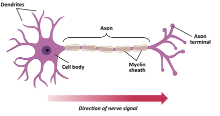
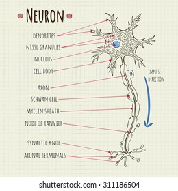
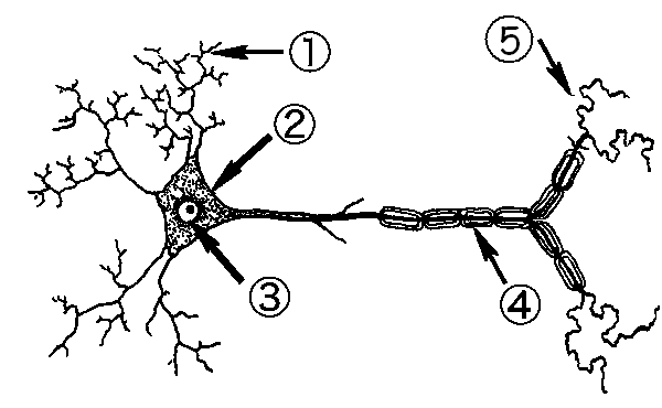
Label the parts of a typical neuron (1, 2, 3, 4, 5, 6) shown in the figure. Structure of Neuron: 1 - Cell body. 2 - Dendrites. 3 - Axon. 4 - Oligodendrocyte. 5 - Myelin sheath. 6 - Synapse ...

Neuron
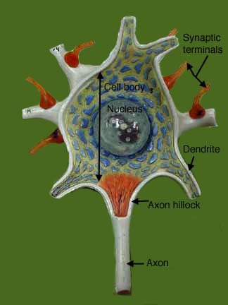
Label Parts of a Neuron Diagram | Quizlet Dendrites. receives impulses from other nerve cells. axon hillock. The cell body...the part of the cell that houses the nucleus and keeps the entire cell alive and functioning. Myelin Sheath. Surrounds the axon an insulates it from surrounding cells and tissues and making signal transitions faster and more efficient. Terminal Buttons.
Multipolar neuron hi-res stock photography and images - Alamy
File:Neuron-no labels.png - Wikimedia Commons ... Diagram of neuron with arrows but no labels. Made using FireFox and GIMP from Neuron.svg by Dhp1080. Date, 1 February 2007, 06:22 (UTC).
566 Neuron labeled Images, Stock Photos & Vectors | Shutterstock
Elsevier Health VerkkoHaluaisimme näyttää tässä kuvauksen, mutta avaamasi sivusto ei anna tehdä niin.
File:Derived Neuron schema with no labels.svg - Wikimedia Commons
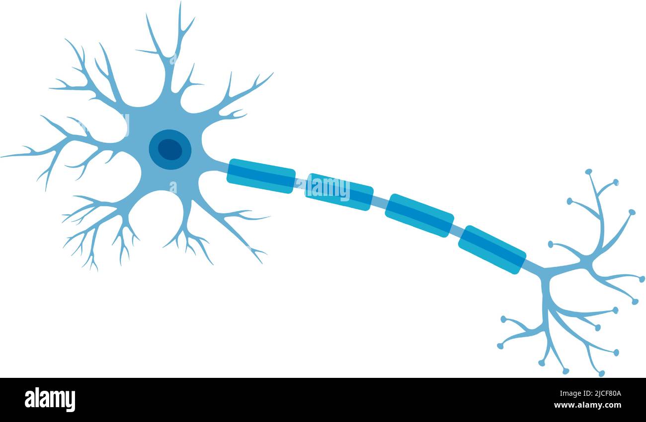
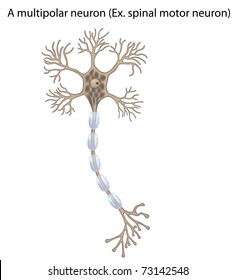
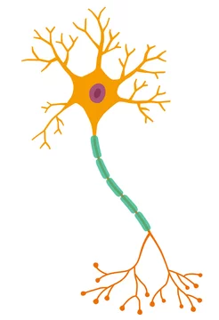
Neuron Diagram & Types | Ask A Biologist - Arizona State University Types of Neurons. There are many types of neurons in your body. Each type is specialized to be good at doing different things. Multipolar neurons have one axon and many dendritic branches. These carry signals from the central nervous system to other parts of your body such as your muscles and glands. Unipolar neurons are also known as sensory ...
Neuron Clip Art, Transparent PNG Clipart Images Free Download ...
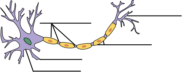
Neuroscience for Kids - Fill In #1 Use the words from the list below to label the following diagram of a neuron in the lines provided. Word Bank. Axon - Cell Body - Dendrites - Myelin Node of ...
Motor Neuron Detailed Accurate Nonlabeled Stock Illustration ...
Neuron Diagram Unlabeled neuron, (1). axon, cell body, dendrites, nucleus, terminal. Unlabeled diagram of a motor neuron (try labeling: axon, dendrite, cell body, myelin, nodes of Ranvier, motor end plate).Read the definitions, then label the neuron diagram below. axon - the long extension of a neuron that carries nerve impulses away from the body of the cell.

Identify the labels of the diagram.
Guide for authors - Cell VerkkoFigure labels Please use uniform lettering and sizing in your original artwork and embed the used fonts if the application you are using provides that option. To ensure labels are legible, we suggest using the following fonts in your illustrations: Arial, Helvetica, Courier, Times, Times New Roman, and/or Symbol, sizing them according to the final figure size.
Neuron B&w Clip Art at Clker.com - vector clip art online ...
Neurons (With Diagram) - Biology Discussion A neuron is a structural and functional unit of the neural tissue and hence the neural system. Certain neurons may almost equal the length of body itself. Thus neurons with longer processes (projections) are the longest cells in the body. Human neural system has about 100 billion neurons. Majority of the neurons occur in the brain.
Labeled Diagram of the Neuron Stock Vector - Illustration of ...
Decoding locomotion from population neural activity in moving C … Verkko29.7.2021 · Red arrows highlight peaks in the temporal derivative of activity of neuron #24 and #110, while cyan arrows highlight peaks of neuron #44. Y- and X-axes labels and scales are preserved within individual rows and columns, respectively. Light green shading indicates the held-out portion of the recording. (d) Same as c but for curvature.
Chapter 13: Nervous Tissue Flashcards | Chegg.com
Understanding AlexNet | LearnOpenCV Verkko13.6.2018 · The second fully connected layer feeds into a softmax classifier with 1000 class labels. ... Because he had memorized the answers to questions covered in the class without understanding the underlying concepts. ... in 2012. In dropout, a neuron is dropped from the network with a probability of 0.5. When a neuron is dropped, ...
New tool offers snapshots of neuron activity | MIT News ...
566 Neuron labeled Images, Stock Photos & Vectors Find Neuron labeled stock images in HD and millions of other royalty-free stock photos, illustrations and vectors in the Shutterstock collection.
The Nervous System Test Yourself - WikiEducator
FitzHugh-Nagumo model - Scholarpedia Jan 27, 2012 · The resting equilibrium of the FitzHugh-Nagumo model shifts slowly to the right, and the state of the system follows it smoothly without firing spikes. In contrast, when the stimulation is increased abruptly, even by a smaller amount, the trajectory could not go directly to the new resting state, but fires a transient spike; see figure.
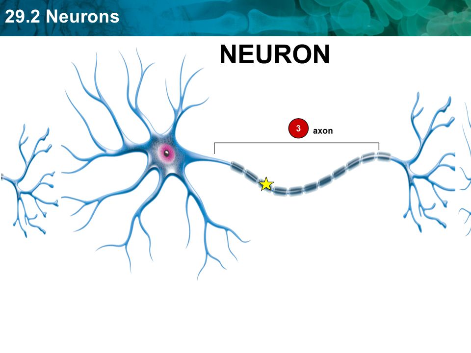
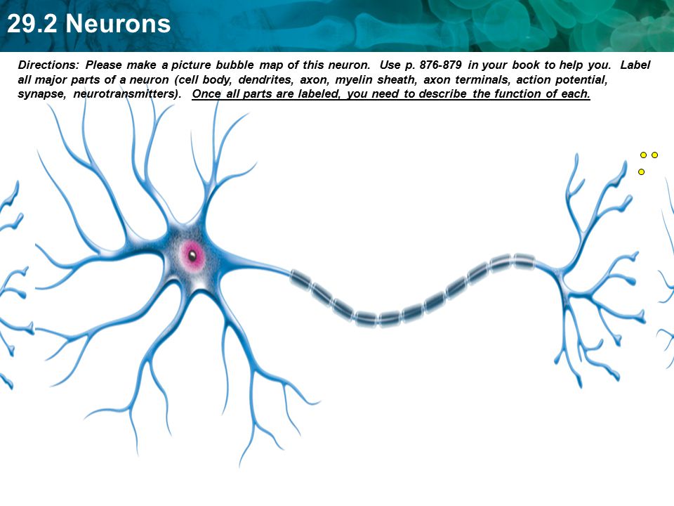
29.2 Neurons Set up Cornell Notes on pg. 39 Topic: 29.2 ...
Picture of a neuron without labels Picture of a neuron without labels 4. in is the label of the input neuron of . A firing rule E=ac → ad ∈ R i can be applied in neuron ˙i if it contains b spikes, b ≥ c, and ab belongs to the language associated to E. For applying rules of type ab=ac → ad, the neuron must contain exactly b spikes.
Neurilemma Images – Browse 33 Stock Photos, Vectors, and ...
Picture of the Skin - WebMD VerkkoWebMD's Skin Anatomy Page provides a detailed image of the skin and its parts as well as a medical definition. Learn about the skin's function and conditions that may affect the skin.
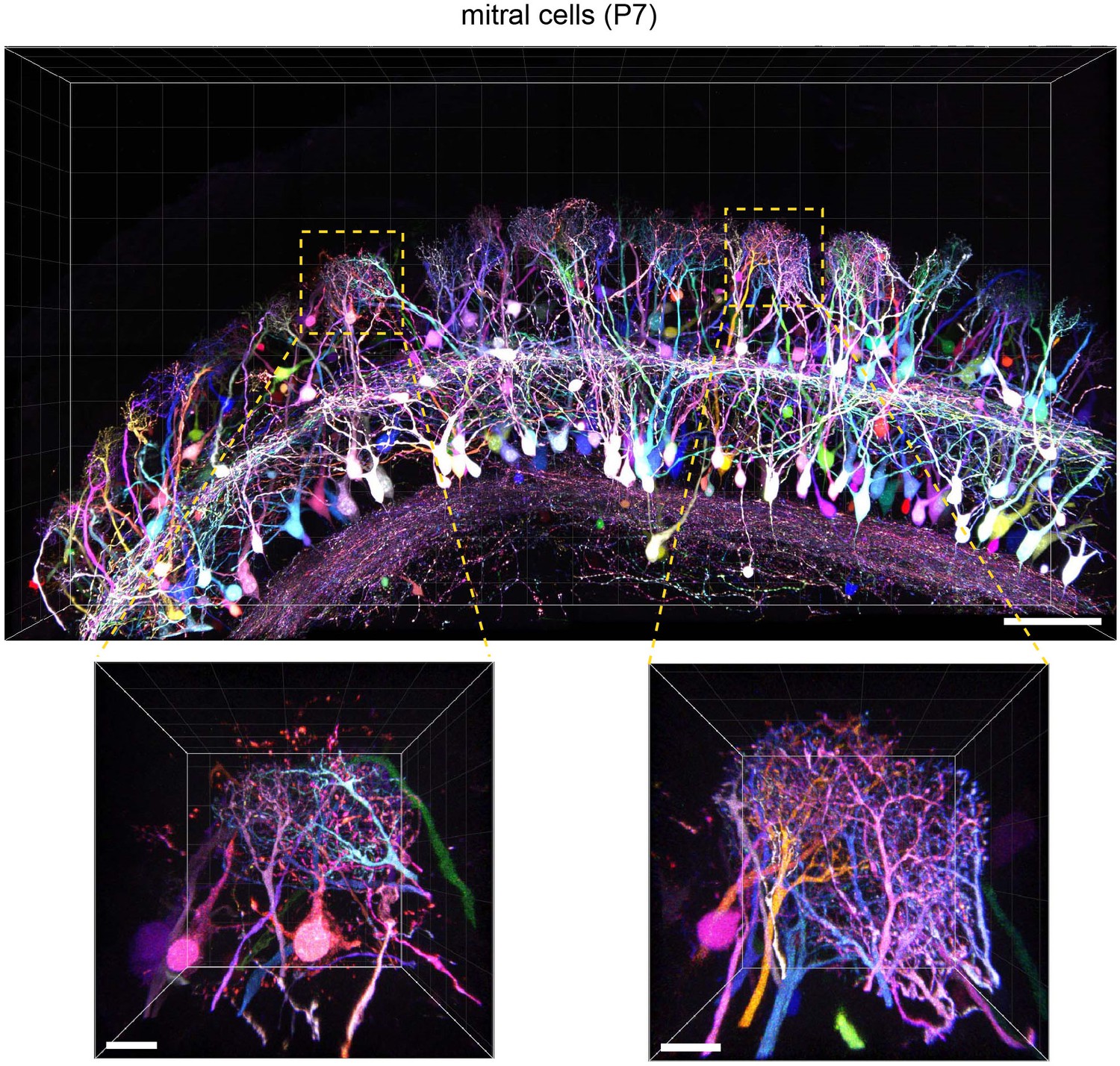
Bright multicolor labeling of neuronal circuits with ...
Photo-labeling neurons in the Drosophila brain - ScienceDirect The cell body of the neuron of interest cannot be located before starting the photo-labeling procedure (step 2 of the photo-label individual neurons section). Potential solution. If the cell body of the neuron of interest is located in the center of the brain, it may be difficult to image.

Neuron Nerve Cell Diagram Blank Sketch Coloring Page ...
MonkeyLogic - Brown University Introduction. MonkeyLogic is a MATLAB toolbox for the design and execution of psychophysical tasks with high temporal precision.It is structured to allow for the flexible construction of sensory, motor, or cognitive tasks that are based upon the interaction of a subject with visual stimuli through the use of eye-position, joystick, button, lever, and / or keyboard input.
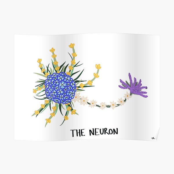
Flowered Neuron Diagram" Poster for Sale by magifur | Redbubble
Picture of a neuron without labels Picture of a neuron without labels alexandra daddario hot one the woman Interneurons connect various neurons within the brain and spinal cord. The simplest type of neural pathway is a monosynaptic (single connection) reflex pathway, like the knee-jerk reflex.
Neuron Models

How to draw Neuron diagram Step by Step for Beginners
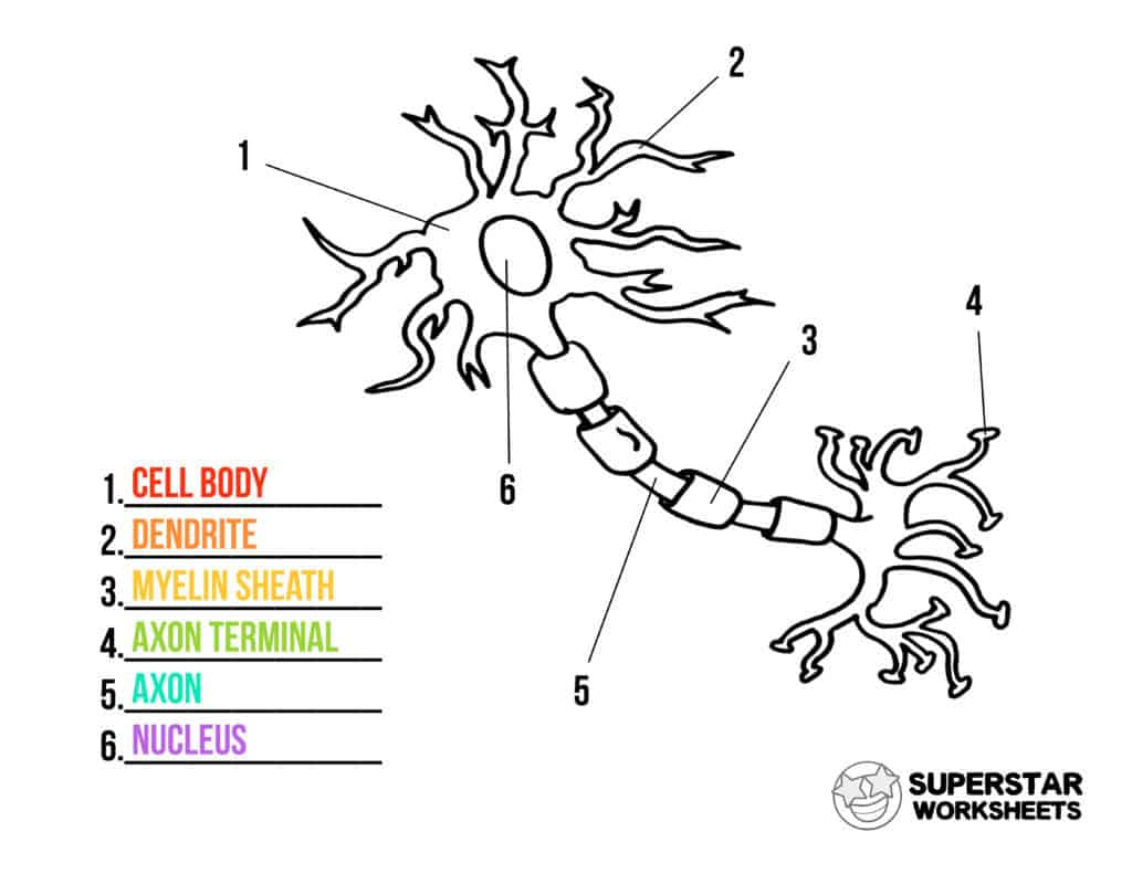
Neuron Cell Worksheets - Superstar Worksheets
The resting potential: introducing foundations of the nervous ...

Labeling a Neuron Quiz

Untitled Document

Free Motor neuron Graphic Vector - Stock by Pixlr
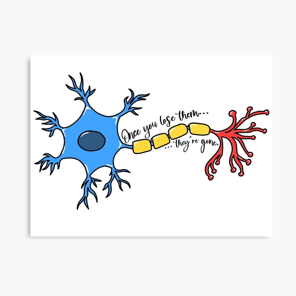
Neurons: once you lose them, they're gone!" Art Board Print ...

Draw the structure of a neuron and explain its function.
Neuroscience for Kids - Fill In #1
Blausen 0657 - Multipolar neuron - English labels | AnatomyTOOL
Label The Neuron Clip Art at Clker.com - vector clip art ...

Label a Neuron Quiz
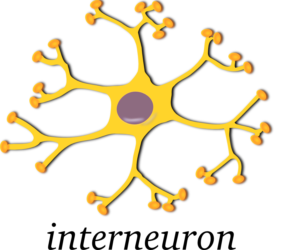
OnlineLabels Clip Art - neuron-interneuron

Sensory Neuron Diagram | Quizlet
Nervous System - Label the Neuron
Nervous System and Neurons - ppt download
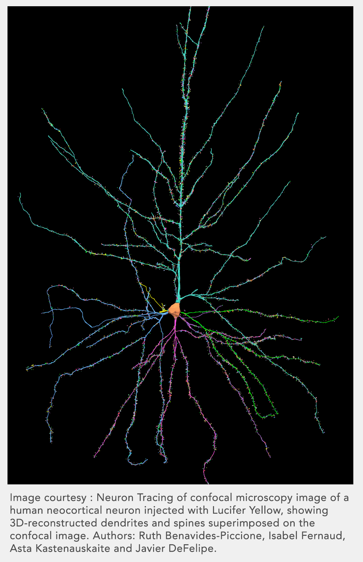
Neuron Reconstruction - MBF Bioscience

Label the parts of a typical neuron (1, 2, 3, 4, 5, 6) shown ...
Post a Comment for "43 picture of a neuron without labels"