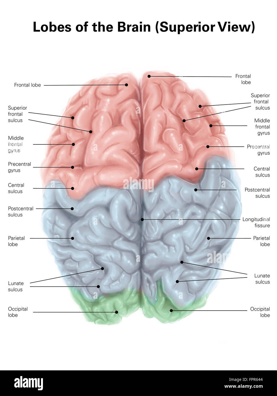
42 brain pictures and labels
Label the Brain Worksheets (SB11585) - SparkleBox Parts of the Body Topic Word Cards (SB275). A set of words with accompanying pictures linked to a topic on parts of the body. Great for classroom displays, word banks or laminating for other topic-related activities. 101 labeled brain images and a consistent human cortical labeling ... 101 labeled brain images and a consistent human cortical labeling protocol Abstract We introduce the Mindboggle-101 dataset, the largest and most complete set of free, publicly accessible, manually labeled human brain images.
Brain Label | Human anatomy and physiology, Basic anatomy and ... Brain Label Image of the brain showing its major features for students to practice labeling. Answers are included. Biologycorner 17kfollowers More information Brain Label Find this Pin and more on Anatomy & Physiologyby Page Johnson. Basic Anatomy And Physiology Brain Anatomy Science Education Physical Science Science Experiments Science Notebooks
Brain pictures and labels
Human Brain Diagram Photos and Premium High Res Pictures - Getty Images 1,018 Human Brain Diagram Premium High Res Photos Browse 1,018 human brain diagram stock photos and images available, or start a new search to explore more stock photos and images. of 17 NEXT Illustration Picture of Brain Anatomy - Brain - eMedicineHealth Medical Illustrations Picture of Brain The brain is the complex organ responsible for processing sensory information (sound, touch, taste, sight, and smell). The brain controls voluntary and involuntary movements. Signals from the brain tell muscles to contract. Input from the brain controls the function of other organs in the body. Drawing Of The Brain With Labels - Painting Valley We collected 36+ Drawing Of The Brain With Labels paintings in our online museum of paintings - PaintingValley.com. ADVERTISEMENT LIMITED OFFER: Get 10 free Shutterstock images - PICK10FREE brain human diagram labeled anatomy label system easy physiology infant coronal neat nervous spinal simple cord rat Brain Diagram Labele... 633x512 41 0
Brain pictures and labels. Brain Label (Remote) - The Biology Corner This brain labeling activity was created for remote learners as an alternative to the labeling and coloring worksheet we would traditionally do in class. Instead of coloring and labeling on printouts, students use google slides to drag labels to the images or type the answers into text boxes. Whole Brain Segmentation and Labeling from CT Using Synthetic MR Images ... With MALP-EM processing of the ground truth T1-w as the reference, we compute the Dice coefficient between multi-atlas segmentations using either the subject CT images with MV label fusion (red), or synthetic T1-w with MV (green) or JLF (blue), as the label fusion, and MALP-EM (yellow).Note that MALP-EM uses the OASIS atlas with manually delineated labels, while the other three use the ... Brain - Human Brain Diagrams and Detailed Information Sensory information is combined, evaluated, and compared to prior experiences, providing the brain with an accurate picture of its conditions. The association areas also work to develop plans of action that are sent to the brain's motor regions in order to produce a change in the body through muscles or glands. Association areas also work to ... Labeling Brain Structures - John Muschelli 1 Labels in template space. In Processing Within-Visit MRI, we registered the T1 image to the Eve template using a non-linear registration (SyN) (Avants et al. 2008). Also, we applied this transformation to the intensity-normalized T1, T2, and FLAIR images, so that these image are located in the same space as the Eve atlases. We can overlay the ...
Parts of the brain: Learn with diagrams and quizzes - Kenhub Labeled brain diagram First up, have a look at the labeled brain structures on the image below. Try to memorize the name and location of each structure, then proceed to test yourself with the blank brain diagram provided below. Labeled diagram showing the main parts of the brain Blank brain diagram (free download!) Label Brain Diagram Printout - EnchantedLearning.com Label the Brain Anatomy Diagram The Brain Read the definitions below, then label the brain anatomy diagram. Cerebellum - the part of the brain below the back of the cerebrum. It regulates balance, posture, movement, and muscle coordination. Corpus Callosum - a large bundle of nerve fibers that connect the left and right cerebral hemispheres. Brain: Anatomy, Pictures, Functions, and Conditions The brainstem is an area located at the base of the brain that contains structures vital for involuntary functions such as the heartbeat and breathing. The brain stem is comprised of the midbrain, pons, and medulla. 3 Midbrain The midbrain is often considered the smallest region of the brain. Brain Image Labeling Using Multi-atlas Guided 3D Fully Convolutional ... To address these limitations, in this paper, we propose a multi-atlas guided 3D fully convolutional networks (FCN) for brain image labeling. Specifically, multi-atlas based guidance is incorporated during the network learning. Based on this, the discriminative of the FCN is boosted, which eventually contribute to accurate prediction.
Parts of the Brain Activity for Kids, Brain Diagram, and Worksheets for ... This brain coloring sheet was my kids' favorite! Brain worksheet for grade 2. Parts of the Brain Worksheet - Label the human brain by writing the number on the brain template; Label the Brain Parts Worksheet - Use the brain vocabulary from the word bank to label the brain areas; Brain Activity for kids 75,682 Brain Anatomy Stock Photos and Images - 123RF Brain Anatomy Stock Photos And Images 75,682 matches Page of 757 Brain lobes vector illustration. Human brain infographic vector. Brain lobes functions Serotonin pathway. Humans brain with serotonin pathways. psychiatric and neurological disorders. 3D render of a medical image showing male figure with brain tumour Neocortex vector illustration. The Brain - Diagram and Explanation BRAIN STEM: The part of the brain that connects to the spinal cord. The brain stem controls functions basic to the survival of all animals, such as heart rate, breathing, digesting foods, and sleeping. It is the lowest, most primitive area of the human brain. CEREBELLUM: Two peach-size mounds of folded tissue located at the top of the brain ... Labeled brain anatomy Images, Stock Photos & Vectors - Shutterstock Find Labeled brain anatomy stock images in HD and millions of other royalty-free stock photos, illustrations and vectors in the Shutterstock collection. Thousands of new, high-quality pictures added every day.
Brain Label - The Biology Corner Image of the brain showing its major features for students to practice labeling. Answers are included.
Nervous System - Label the Brain This brain part controls thinking. This brain part controls balance, movement, and coordination. This brain part controls involuntary actions such as breathing, heartbeats, and digestion. This part of the nervous system moves messages between the brain and the body. This part of the cerebrum interprets and sorts information from the senses.
How to Draw a Brain: 14 Steps (with Pictures) - wikiHow Once you've drawn the cartoon or realistic brain, you can go back and add color or label the parts. Method 1 Sketching an Easy Cartoon Brain 1 Draw a large bean shape to make the outline for the brain. Use a pencil to sketch a kidney bean shape on your paper. You can make the outline for the brain any size you like.

Cigarette Warning Labels With Ugly Pictures Deliver Anti-Smoking Message Better Than Text-Only ...
Diagram of the Brain and its Functions - Bodytomy Given below is a labeled diagram showing the brain stem and its related structures. Brain Stem and Structures Cerebellum The word 'cerebellum' literally means little brain. It is the second largest part of the brain, and is located at the back, below the occipital lobe, beneath the cerebrum and behind the brain stem.
Human Brain Photos and Premium High Res Pictures - Getty Images Browse 27,835 human brain stock photos and images available, or search for human brain anatomy or human brain illustration to find more great stock photos and pictures. Related searches: human brain anatomy. human brain illustration.
101 Labeled Brain Images and a Consistent Human Cortical Labeling ... In this article we introduce this dataset of manually edited brain image labels applied to the T1-weighted MR images of publicly available multi-modal data acquired from healthy individuals. We also introduce a benchmark for the evaluation of automated registration/segmentation/labeling methods by comparing the manual labels according to this "Desikan-Killiany-Tourville" (DKT) protocol with automatically generated labels.
Whole Brain Segmentation and Labeling from CT Using Synthetic MR Images W e use MALP-EM [9] to provide whole-brain segmentation and labeling from the synthetic MR images. Since the synthetic MR images are naturally registered with the CT images, the result is a ...
Picture of the Brain - WebMD • The cerebellum is at the base and the back of the brain. The cerebellum is responsible for coordination and balance. The brain is also divided into several lobes: • The frontal lobes are...

Label the Brain Worksheets (SB11585) - SparkleBox | Human brain diagram, Brain diagram, Human brain
Brain: Atlas of human anatomy with MRI - e-Anatomy - IMAIOS MRI Atlas of the Brain. This page presents a comprehensive series of labeled axial, sagittal and coronal images from a normal human brain magnetic resonance imaging exam. This MRI brain cross-sectional anatomy tool serves as a reference atlas to guide radiologists and researchers in the accurate identification of the brain structures.
Drawing Of The Brain With Labels - Painting Valley We collected 36+ Drawing Of The Brain With Labels paintings in our online museum of paintings - PaintingValley.com. ADVERTISEMENT LIMITED OFFER: Get 10 free Shutterstock images - PICK10FREE brain human diagram labeled anatomy label system easy physiology infant coronal neat nervous spinal simple cord rat Brain Diagram Labele... 633x512 41 0
Illustration Picture of Brain Anatomy - Brain - eMedicineHealth Medical Illustrations Picture of Brain The brain is the complex organ responsible for processing sensory information (sound, touch, taste, sight, and smell). The brain controls voluntary and involuntary movements. Signals from the brain tell muscles to contract. Input from the brain controls the function of other organs in the body.
Human Brain Diagram Photos and Premium High Res Pictures - Getty Images 1,018 Human Brain Diagram Premium High Res Photos Browse 1,018 human brain diagram stock photos and images available, or start a new search to explore more stock photos and images. of 17 NEXT


Post a Comment for "42 brain pictures and labels"